In this article:
What is an ECG?
When is an ECG used?
How an ECG is carried out
Types of ECG
Getting your results
Are there any risks or side effects?
What is an ECG?
When is an ECG used?
How an ECG is carried out
Types of ECG
Getting your results
Are there any risks or side effects?
Note: the information below is a general guide only. The arrangements, and the way tests are performed, may vary between different hospitals. Always follow the instructions given by your doctor or local hospital.
What is an ECG?
Your heart produces tiny electrical impulses which spreads through special routes (fibres) in the heart muscle to make the heart contract. These impulses can be detected by an electrocardiogram (ECG) machine. An electrocardiogram (ECG) is the recording of this electrical activity generated by your heart. You may have an ECG to help find the cause of symptoms such as the feeling of a 'thumping heart' (palpitations) or chest pain. Sometimes it is done as part of routine tests - for example, before you have an operation.The ECG machine records electrical impulses coming from your body - it does not put any electricity into your body. Therefore, the ECG test is painless and harmless.
 |
| Schematic diagram of normal sinus rhythm for a human heart as seen on ECG. Credit: Agateller (Anthony Atkielski) / Public domain |
 |
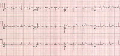
| A normal ECG tracing. Credit: Queen's University School of Medicine |
Despite having a similar name, an ECG is not the same as an echocardiogram, which is a scan of the heart.
When is an ECG used?
An ECG can be used to investigate symptoms of a possible heart problem, such as chest pain, sudden noticeable heartbeats (palpitations), dizziness and shortness of breath. It is often used alongside other tests to help diagnose and monitor conditions affecting your heart.An ECG can help detect the following heart disorders:
- Abnormal heart rhythms (arrhythmias). If the heart rate is very fast, very slow, or irregular. There are various types of irregular heart rhythm with characteristic ECG patterns.
- Coronary heart disease. Where the heart's blood supply is blocked or interrupted by a build-up of fatty substances.
- A heart attack (myocardial infarction) and if it was recent or some time ago. A heart attack causes damage to heart muscle and it heals with scar tissue. These can be detected by abnormal ECG patterns.
- Cardiomyopathy. Where the heart walls become thickened or enlarged from certain types of diseases affecting the heart.
- An enlarged heart (cardiomegaly). Basically, this causes bigger impulses than normal.
How an ECG is carried out
There are several different ways an ECG can be carried out. Generally, the test involves attaching a number of small, sticky sensors called electrodes to your arms, legs and chest to detect electrical impulses coming from different directions within your heart. The electrodes are connected by wires to an ECG recording machine. The machine detects and amplifies the electrical impulses that occur at each heartbeat and records them on to a paper or computer. A few heartbeats are recorded from the different sets of electrodes. The test takes about five minutes to do.Types of ECG' below).
You do not need to do anything special to prepare for an ECG test. You can eat and drink as normal beforehand.
Before the electrodes are attached, you will usually need to remove your upper clothing, and your chest may need to be shaved or cleaned. Once the electrodes are in place, you may be offered a sheet to cover yourself.
The test itself usually only lasts a few minutes, and you should be able to go home soon afterwards or return to the ward if you're already staying in hospital.
Types of ECG
There are 3 main types of ECG:- a resting ECG – carried out while you're lying down in a comfortable position
- a stress or exercise ECG – carried out while you're using an exercise bike or treadmill
- an ambulatory ECG – the electrodes are connected to a small portable machine worn at your waist so your heart can be monitored at home for 1 or more days
For example, an exercise ECG may be recommended if your symptoms are triggered by physical activity, whereas an ambulatory ECG may be more suitable if your symptoms are unpredictable and occur in random, short episodes.
Getting your results
An ECG recording machine will usually show your heart rhythm and electrical activity as a graph displayed electronically or printed on paper (see images above).For an ambulatory ECG, the ECG machine will store the information about your heart electronically, which can be accessed by a doctor when the test is complete.
Although the ECG machine prints out the result at the end of the test, you may not be able to get the results of your ECG immediately. This is because the recordings may need to be looked at by a specialist doctor to see if there are signs of a potential problem. Other tests may also be needed before it is possible to tell you whether there is a problem.
You may need to visit the hospital, clinic or your healthcare center few days later to discuss your results with a doctor.
Are there any risks or side effects?
An ECG is a quick, safe and painless test. No electricity is put into your body while it's carried out.There may be some slight discomfort when the electrodes are removed from your skin – similar to removing a sticking plaster – and some people may develop a mild rash where the electrodes were attached.
An exercise ECG is performed under controlled conditions. The person carrying out the test will carefully monitor you, and they’ll stop the test if you experience any symptoms or start to feel unwell.
The British Heart Foundation and the US Department of Health have more information about what an exercise ECG involves.
Reference(s)
1). National Heart Lung and Blood Insitute (NIH): Electrocardiogram. Available online: https://www.nhlbi.nih.gov/health-topics/electrocardiogram
2). British Heart Foundation: Electrocardiogram (ECG). Available online: https://www.bhf.org.uk/informationsupport/tests/ecg


No comments:
Post a Comment
Got something to say? We appreciate your comments: